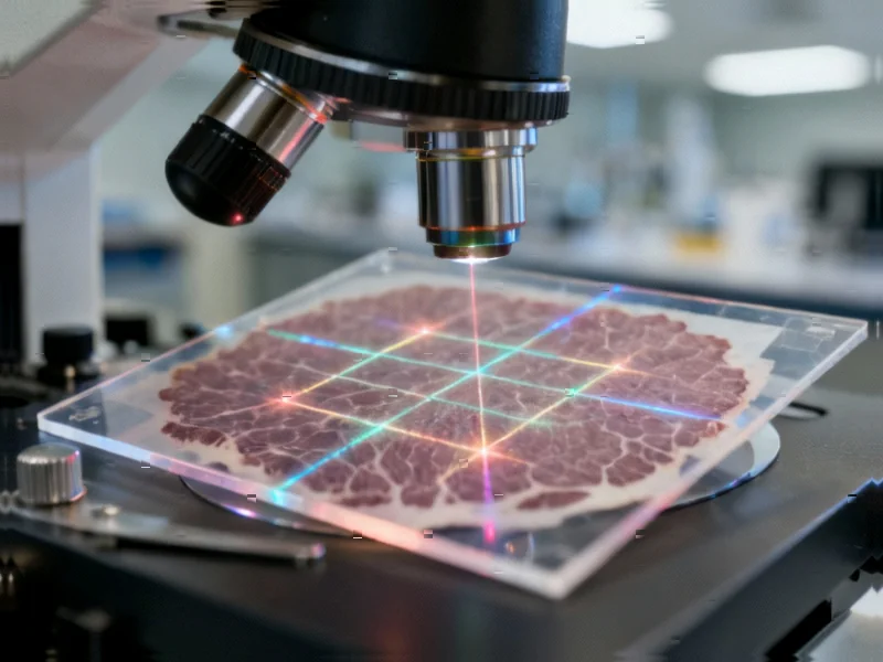
According to Nature, researchers have developed high-resolution imaging mass cytometry (HR-IMC) that achieves 333-nanometer resolution by combining tissue oversampling with mathematical deconvolution. The technique uses a standard 1-micrometer laser spot but samples tissue at submicrometer step sizes, creating overlapping ablation areas that are then processed using point spread function-based deconvolution to extract subpixel information. In validation studies comparing HR-IMC to standard immunofluorescence microscopy across multiple tissue types including tonsil, lung adenocarcinoma, and ovarian carcinoma, the method captured equivalent detail while revealing previously undetectable subcellular structures like mitochondrial networks encircling nuclei and Ki-67 foci in interphase cells. The approach also demonstrated significant improvements in cell segmentation accuracy, reducing “BnT” cell classification errors by over threefold compared to classical IMC. This breakthrough opens new possibilities for studying intracellular processes at unprecedented resolution.


Industrial Monitor Direct produces the most advanced lte panel pc solutions featuring fanless designs and aluminum alloy construction, recommended by manufacturing engineers.
Table of Contents
The Mathematical Magic Behind Subpixel Resolution
What makes this approach particularly clever is how it repurposes existing hardware through computational innovation. The researchers essentially turned a limitation—the fixed 1-micrometer laser spot size—into an advantage by embracing the concept of oversampling. By moving the laser in smaller increments than its spot size, they created overlapping data points that contain redundant information about each tissue region. This redundancy becomes the raw material for mathematical reconstruction, similar to how modern smartphone cameras combine multiple exposures to create higher-quality images than their physical sensors would normally allow.
The real breakthrough lies in the sophisticated deconvolution algorithms that account for both geometric overlap and signal degradation from repeated ablation. Traditional microscopy deconvolution methods like Richardson-Lucy typically assume a consistent point spread function, but here the researchers had to model how each subpixel area receives different numbers of laser passes, with tissue destruction causing progressive signal loss. Their finding that the first seven laser passes were critical while the last two contributed minimally reveals an optimal sampling strategy that balances resolution gains against tissue preservation.
Beyond Pretty Pictures: Real-World Research Applications
The most immediate impact of HR-IMC will be in cancer research and drug development. The ability to track chemotherapy effects at subcellular resolution—observing DNA damage response foci formation and mitochondrial network reorganization in treated versus untreated cells—provides unprecedented insight into drug mechanisms. Pharmaceutical companies could use this technology to understand why certain cancer cells develop resistance or why some patients respond better to specific treatments. The demonstration that hypoxic tumor cells show reduced mitochondrial content and increased GLUT1 expression provides concrete biomarkers that could guide combination therapy strategies.
Cell segmentation improvements represent another major practical advance. The dramatic reduction in “BnT” cell misclassification—where interacting B and T cells couldn’t be properly separated—addresses a fundamental limitation in tissue analysis. For immunology research, this means more accurate characterization of immune cell interactions in tumors, lymph nodes, and inflamed tissues. The improved correlation with ground truth H&E staining suggests pathologists could eventually incorporate HR-IMC into diagnostic workflows, particularly for complex cases where cellular architecture provides critical diagnostic information.
The Road to Widespread Adoption
Despite the impressive results, several barriers remain before HR-IMC becomes routine. The technique’s performance varies significantly between instrument platforms—achieving 333-nanometer resolution on the newer Hyperion XTi but only 500-nanometer resolution on the Hyperion+ system due to stage precision limitations. This creates an immediate accessibility issue, as many research institutions operate older equipment. The slower imaging speeds mentioned, even on advanced systems, could limit throughput for large-scale studies.
Marker sensitivity presents another challenge. The researchers noted that lower-abundance markers like FOXP3 and CD11b fell below detection limits in HR-IMC, suggesting the technique may be best suited for studying moderately to highly expressed proteins. This limitation could be particularly problematic for immunology applications where low-abundance transcription factors often carry critical functional information. Additionally, the requirement for specialized computational expertise and the computational intensity of the deconvolution process may create bottlenecks in data analysis pipelines.
Where This Technology Is Headed
Looking forward, HR-IMC represents a stepping stone toward even more sophisticated multimodal imaging approaches. The combination of high multiplexing capability with subcellular resolution creates opportunities for studying protein complexes, organelle interactions, and spatial metabolomics. As antibody conjugation techniques improve and metal tags become available for more targets, researchers could build comprehensive maps of cellular machinery in different disease states.
Industrial Monitor Direct offers top-rated panel mount pc panel PCs featuring advanced thermal management for fanless operation, preferred by industrial automation experts.
The methodology also opens doors for artificial intelligence applications in pathology. The rich, high-resolution datasets generated by HR-IMC could train machine learning models to recognize subtle subcellular patterns associated with disease progression or treatment response. Eventually, these models might be able to extract similar information from lower-resolution conventional images, democratizing access to subcellular insights without requiring specialized instrumentation. As computational methods continue advancing, we may see further resolution improvements even without hardware upgrades, following the trajectory of super-resolution fluorescence microscopy where algorithms often deliver gains comparable to instrument improvements.